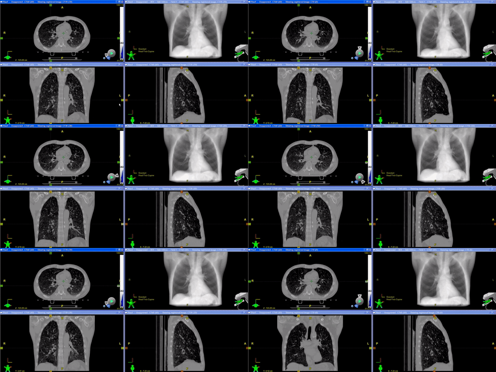
The treatment for lung cancer using Cyberknife often requires the fiducial markers implanted in the patient’s lung to move with the patient’s breathing. The purpose of this study is to observe whether the patient’s tumor also moves alongside with the fiducials and evaluate if the treatment is effective with proper margins. This study consisted of 18 upper lung cancer patients, 8 lower lung cancer patients, and 1 middle lung cancer patient. The fiducial markers were first tracked through Cyberknife’s CT program. Then the 4D CT contours of each patient were used to find the center of mass of the tumor. Finally, the center of mass and the fiducial marker movements were compared. The centroids of the fiducial markers were used as the coordinates for comparison. The root mean square error was found for the superior and inferior movements of the tumor for comparison to the fiducial markers. The root mean square error, which shows the statistical spread of the compared values, was found for the superior and inferior movements of the tumor for comparison to the fiducial markers. The values of the lower and upper lung patients were separated to see if there was a significant difference in tracking from where the tumor was regarding the diaphragm movement. The upper lung values gave three outliers that were out of the range of below 2mm. The patients with lower lung tumors had more spread values in comparison to the upper lung tumor patients. The results showed a variation of the average spread between the tumor and fiducial movements, which most were relatively small, but some had significant discrepancies. The discrepancies may be from the markers not staying in sync with the patients’ breathing, but they also could be from the markers not being tracked well from being shadowed by each other. Future research can develop and lower the uncertainty of fiducial marker tracking of lung tumors.

(Source: Aquilion LB 4D CT, https://youtu.be/1uBkvOyp1b8 )